
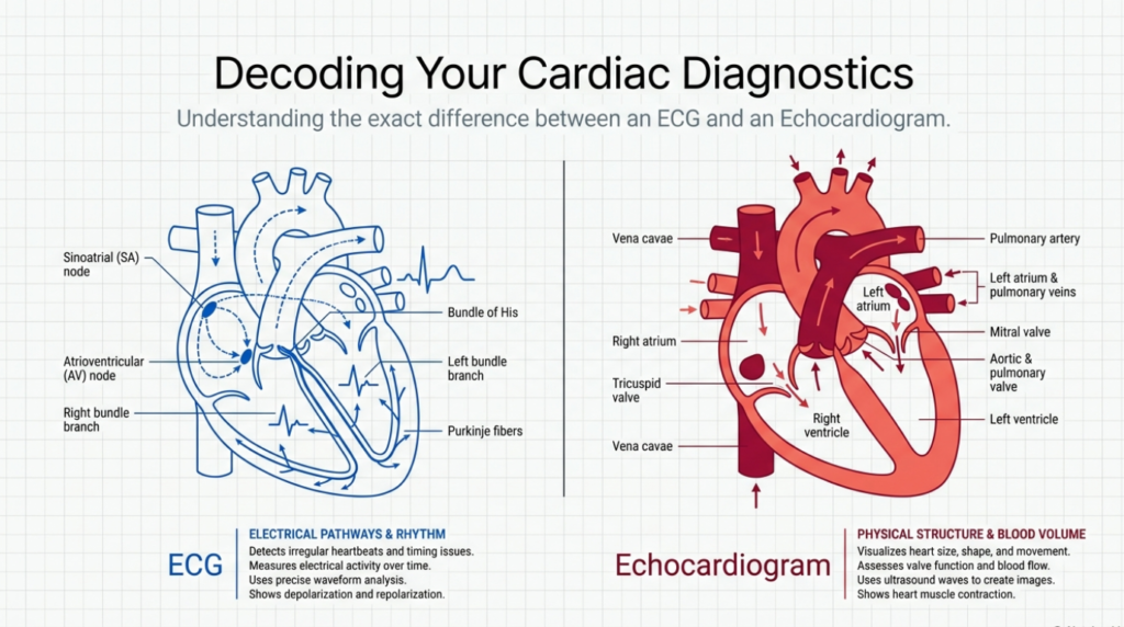
When your physician suspects heart palpitations, chest pain, shortness of breath, dizziness, fatigue, fainting or an abnormal heart sound, you may be asked to have an electrocardiogram test, an echocardiogram or both. They are different cardiac diagnostic tests that examine different aspects of your heart function.
An ECG is a test that traces your heart’s electrical activity. An echocardiogram uses ultrasound imaging to look at your heart. One is not a replacement for the other. The right test, or tests, will be chosen based upon your symptoms.
If you are experiencing possible heart-related symptoms, you don’t want to guess at which test, or combination of tests, to have. Your family doctor Etobicoke can evaluate and advise you based upon your symptoms, medications, risk factors and heart exam.
When you need a timely assessment of possible new symptoms but you don’t have a regular family doctor’s availability, you might want to consider going to an urgent care clinic Etobicoke for an initial evaluation, which may include ECG, echocardiogram, further testing or referrals based upon their findings.
Some key points to know about ECGs and echocardiograms:
An ECG traces the electrical impulses of the heart.
An echocardiogram uses ultrasound to see the heart.
A standard ECG takes only a few minutes to record.
A standard transthoracic echocardiogram usually takes about 30 to 40 minutes.
Neither test usually exposes you to radiation.
A normal ECG does not always mean that your heart structure is normal.
A normal echocardiogram does not always mean that your heart rhythm is normal.
Some people benefit from having both tests, since the tests address different concerns.
ECG Overview and What it Measures
An electrocardiogram, abbreviated as ECG or EKG, is a test that measures your heart’s electrical activity. Electrodes, small sticky patches that record the electricity, are placed on your chest. They may also be placed on your legs and wrists, depending upon what your doctor wants to assess. The machine traces this electricity and displays or prints it as a graph. The ECG machine records the electricity; it does not send electricity to your heart.
Each heartbeat begins with an electrical impulse. The heart’s conduction system carries the impulse to the heart’s chambers in a specific pattern. Each chamber contracts, causing the heart to beat and pump blood. An ECG can help your doctor determine your heart rate or rhythm, identify abnormal heart rhythms, recognize conduction problems and assess the likelihood that you are having a heart attack.
Your doctor may order an ECG to help answer a number of questions, such as: Why are you experiencing palpitations or skipped beats? Why are you feeling lightheaded or faint? Why do you have chest discomfort or why are you short of breath? Why does your blood pressure or pulse seem abnormal? Why are you experiencing fatigue with minimal exertion? Does the ECG show evidence of a heart attack, rhythm problems or heart strain?
Different kinds of ECG tests:
Your doctor may recommend a resting ECG, the standard type of ECG. The test is typically quick and simple. You’ll lie on a table while your heart rate and rhythm are recorded for a short period of time. Only a few minutes of the test are actually spent recording the electrical impulses. You must keep your body still while the test is in progress, since muscle contractions or shivering can affect the ECG tracing.
A standard ECG provides only a small sample of your heart’s electrical activity. If you are experiencing intermittent palpitations or skipped beats, you doctor may suggest using a Holter monitor or another type of ECG that monitors your heart’s electrical activity continuously over a period of time.
An exercise ECG, also called an exercise stress test, looks at your heart’s electrical activity while you are walking on a treadmill. An ECG can be tailored to your individual level of fitness and your doctor’s concerns. Your doctor considers many factors when recommending an ECG, such as your symptoms, physical exam, medical history and test results. There is more information about ECGs in this overview of electrocardiography.
Echocardiogram Overview and What it Shows
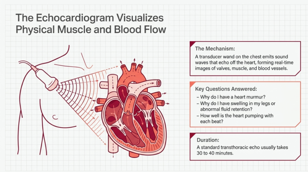
An echocardiogram, sometimes called an echo, is a type of ultrasound test that looks at your heart. This procedure uses sound waves to create images of the heart. A small wand called a transducer is placed on your chest. This emits sound waves which echo off of your heart. These echoes are used to form images of your heart, allowing the doctor to see your heart valves, muscle, chambers and blood vessels.
Unlike an ECG, which looks at your heart’s electricity, an echo can evaluate your heart’s structure and how well your heart is pumping. As the heart muscle tightens and relaxes, the echoes can show how well your heart valves open and close and how much blood your heart is pumping with each beat. This is also helpful in looking at problems with the heart’s blood vessels.
An echocardiogram may be ordered to help your doctor answer the following questions: Why do I have a heart murmur? Why do I feel short of breath, especially with activity? Why do I have swelling in my legs? Does this heart murmur suggest heart disease? Why do I have heart failure symptoms such as fatigue or fluid retention? Why do I have congenital or acquired heart disease or a heart attack?
Different kinds of echocardiograms:
The most common echocardiogram is a transthoracic echocardiogram. You will typically be lying on your back or on your left side on an exam table. Your doctor or a sonographer will apply some gel to your skin, and then will place a transducer over different areas of your chest to capture images of your heart. You may be asked to turn onto your left side or to hold your breath for certain views.
A standard transthoracic echocardiogram usually takes about 30 to 40 minutes. Another type of echocardiogram is called a stress echocardiogram. This looks at how well your heart is pumping at rest and after an activity, such as walking on a treadmill or receiving a medication that makes your heart beat faster. A transesophageal echocardiogram is another type of echo that looks at the heart structure more closely. Your doctor will discuss the specific test with you, and review the preparation and risks that may be associated with the echo that he or she recommends.
Echocardiogram vs ECG: The Main Differences
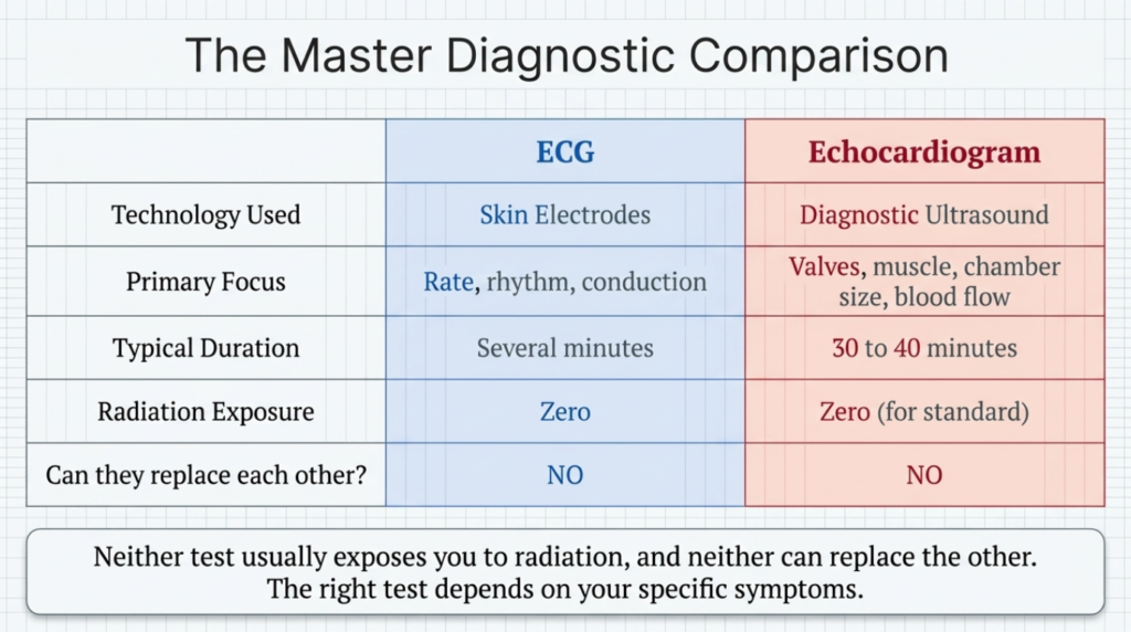
The main difference is that an ECG looks at your heart’s electrical activity and an echocardiogram looks at your heart structure. An ECG can look at the rate and rhythm of the heartbeat, but an echo can show how well the heart is pumping. An ECG and an echo are both important tests and can provide vital information, but they examine different aspects of heart function.
Here is a quick comparison:
ECG Echo
Electrocardiogram Echocardiogram
Uses skin electrodes Diagnostic ultrasound
Focuses on electricity Focuses on structure
Looks at rate and rhythm Assesses valves, muscle, chambers, blood flow
Typical duration: several minutes Several minutes to 30 to 40 minutes for a standard echo
No radiation No radiation for a standard echo
Used for: abnormal heart rhythms Valves, blood flow, function
Can it replace the other test? No No
For example: A patient who has intermittent heart palpitations may need an ECG or a Holter monitor since the main concern is an abnormal heart rhythm. A patient with a new heart murmur and concerns about heart failure may need an echocardiogram.
When a heart attack is suspected, the ECG becomes an essential tool. It is used along with heart tests and blood tests, such as troponin testing. An echo may also be used, since it can look at how well the heart is pumping and whether there are any abnormalities in the heart muscle. However, an echo cannot show the site of a heart attack directly, while an ECG can.
The tests may be used together. For example, in a patient with suspected heart failure, the ECG can look at the rhythm and help determine whether there is a heart attack or conduction system disease. The echo can look at the heart valves, pumping and chamber sizes. The ECG findings might suggest the cause of heart failure, or may appear completely normal if the heart failure is due to valve disease. The two tests are both important, and should be considered together.
Which Heart Test Might You Need?
The choice of test, or tests, depends upon the questions that your doctor is trying to answer and your general condition. Palpitations, an irregular pulse, dizziness or fainting or conduction disease are concerns addressed by an ECG. An echocardiogram may be considered for a heart murmur, shortness of breath, abnormal fluid retention or symptoms of heart failure, or existing or suspected heart disease.
You both tests, along with other cardiac tests, blood tests, X-rays, stress tests or a referral to a cardiologist, may be recommended for certain patients. A discussion with your family doctor Etobicoke is important to decide which tests are indicated.
During a resting ECG, the electrodes are placed on your chest with little or no discomfort. You will need to remain still while the test is being done, but you are not otherwise restricted in any way. You don’t need to prepare for the test in any special way, other than keeping your skin clean and free of lotions, oils or other substances that may interfere with the adhesion of the electrodes.
You can talk to your doctor about medicines you are taking, or you are considering, but you should never stop taking a medication unless your doctor recommends it. For most routine echocardiograms, you can eat or drink as you normally would, or take your medications with plenty of water, unless your test requires special preparation. For example, you may be instructed to fast before a transesophageal echo.
What to expect after a test and how results help guide treatment
An abnormal test result does not always mean that you have a specific disease, just as a normal test result does not always rule out a particular concern. With regard to the ECG, the results might reflect the effects of medications, or might be completely normal despite your symptoms. On the other hand, an ECG might suggest that you are having a heart attack, or that you have conduction abnormalities that require further evaluation. An echocardiogram might also show normal findings despite your symptoms; or, it might identify valve abnormalities, heart muscle problems or fluid around the heart. The results need to be interpreted in detail by your doctor.
Your family doctor Etobicoke can review all of the test results and discuss next steps, including possible medication changes, additional testing, referrals to other doctors or heart therapy recommendations. If you find yourself needing medical attention but you don’t have an appointment with your regular doctor, you can always visit with an urgent care doctor Etobicoke. You don’t want to ignore your symptoms just because you don’t have a family doctor’s appointment available at the time.
When Heart Symptoms Require Emergency Care
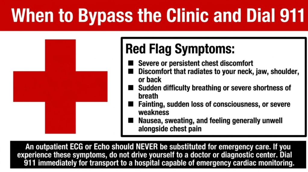
An outpatient ECG or echocardiogram should not be substituted for emergency care. Dial 911 for severe chest discomfort that might suggest a heart attack, especially if you have additional concerns, such as difficulty breathing or fainting. Seek emergency care for severe or persistent chest discomfort, sudden difficulty breathing, fainting or loss of consciousness, severe weakness or if you feel that you may be experiencing a heart attack.
Symptoms of a heart attack may also include sweating, nausea, chest discomfort that radiates to your neck, jaw, shoulder or back and feeling generally unwell. If you are experiencing heart attack-like symptoms, never drive yourself to the nearest diagnostic center or doctor’s office – instead, call for emergency medical assistance. A heart attack typically requires an ECG, heart testing and cardiac monitoring while being transported to an appropriate hospital.
This article discusses electrocardiogram (EKG or ECG) and echocardiogram (echo) testing, their differences, similarities, indications, preparation and possible next steps after receiving abnormal results. The ECG looks at the heart’s electrical activity and the echo uses ultrasound to look at the heart structure. Both tests are important, but they typically address different concerns. The discussion with your family doctor Etobicoke is critical in choosing the right test or tests for your personal situation.
However, the tests are not interchangeable, so a normal ECG does not rule out heart disease, nor does a normal echo rule out an abnormal heartbeat. You should not rely on a normal test result to assure you that everything is fine, since test results should always be interpreted in the context of the overall clinical picture. If your symptoms persist despite a normal ECG or echo, talk to your doctor to determine why your symptoms might be occurring and what further tests or treatment options are available.
Frequently Asked Questions
1. Is an ECG the same as an EKG?
Yes, an EKG and an ECG are the same test. The difference is in the spelling, with ”ECG” being short for electrocardiogram and ”EKG” coming from the German equivalent, Elektrokardiogramm.
2. Is an echocardiogram more accurate than an ECG?
There is no definitive answer to that question, since each test is designed to look at different aspects of heart function. An ECG is more useful for looking at heart rhythm and conduction problems, while an echocardiogram is more useful for looking at heart structure and function.
3. Can an ECG detect blocked arteries?
An ECG can sometimes suggest that you may have reduced blood flow to the heart, or may reveal abnormal findings that suggest that you are having a heart attack. However, ECG findings alone are not enough to diagnose blocked arteries. Additional tests, such as cardiac CT angiography, a standard angiogram or a heart MRI may be needed.
4. Can an echocardiogram detect a heart attack?
An echocardiogram can sometimes identify areas of the heart that are not pumping normally, and it can help evaluate your heart for damage or complications following a heart attack. However, an ECG is typically needed to confirm that you are having a heart attack, as well as additional blood tests that can help your doctor determine the extent of the damage.
5. Which test is better for palpitations?
An ECG is more helpful in evaluating palpitations since they are usually related to abnormal heart rhythms. If your palpitations are intermittent, you may need to wear a Holter monitor or other similar device for a period of time. An echocardiogram may also be needed to look at your heart structure for other underlying heart disease.
6. Which test is better for a heart murmur?
An echocardiogram is more useful for evaluating a heart murmur, since it looks at the anatomy and function of the heart valves. An ECG may also be performed to look at heart rhythm and heart size.
7. Do ECGs or echocardiograms use radiation?
No, a standard ECG does not use radiation, since it looks at the electrical activity of the heart. An echocardiogram also does not use radiation, since it uses ultrasound to look at the heart.
8. Do I need to fast before either test?
Fasting is not usually necessary for a routine ECG or a standard echocardiogram. You may need to fast for a transesophageal echocardiogram, or for an echocardiogram performed with medication, or if you are being sedated for either test. You should follow the instructions provided by your physician or the facility where you are having the test.
9. Can both tests be normal even when symptoms are present?
Yes, it is possible that an ECG or an echocardiogram can be normal despite your symptoms. This could happen for several reasons. For an ECG, a brief test may miss intermittent abnormal heart rhythms, or the ECG findings may be completely normal even when you are experiencing palpitations or other concerns.
On the other hand, an echocardiogram may also appear completely normal, especially if your symptoms are related to an irregular heart rhythm. Even when both tests are normal, your symptoms should still be evaluated carefully, especially if you are experiencing chest discomfort, palpitations or fainting.
10. When should chest symptoms be treated as an emergency?
You should treat chest discomfort as an emergency if it is severe or persistent, especially if you are experiencing additional symptoms, such as difficult breathing, fainting, sudden loss of consciousness or weakness. You should also treat chest discomfort as an emergency if you believe you may be having a heart attack.
Symptoms of a heart attack may include chest discomfort that radiates to your arms, neck, jaw, back or shoulders, nausea, vomiting, sweating, shortness of breath or feeling generally ill. If you are experiencing these or similar symptoms, you should call 911 or ask someone to take you to the nearest emergency room. A heart attack usually requires an ECG, additional heart tests and special care in the hospital, so it is important that you receive emergency care as soon as possible.