
Heart symptoms are varied and frequently non-specific. Shortness of breath, fatigue, diminished exercise tolerance, peripheral edema, palpitations, syncope and chest discomfort can raise concern about cardiac disease, but similar presentations can arise from pulmonary, metabolic, musculoskeletal or other causes. A physician’s timely consideration of diagnostic testing can help differentiate among these possibilities, guiding treatment and management decisions in cases where structural or functional cardiac disease is suspected.
An echocardiogram is a valuable test that enables doctors to evaluate the heart’s structure and function. This procedure can help determine the underlying cause of a patient’s symptoms and allow for faster triage, specialist evaluation or treatment if indicated.
An Overview of Echocardiography
Echocardiography makes use of ultrasound (high-frequency sound waves) to create images of the heart. These visual aids enable physicians to assess the size and shape of the heart, the function of the heart valves and the movement of the heart’s chambers. Unlike an ECG, which records the heart’s electrical activity, an echocardiogram provides information about the heart’s anatomy and physiology.
The availability of echocardiography makes it possible for patients to receive expeditious care and for physicians to obtain crucial diagnostic information earlier in the clinical decision-making process. Not every heart-related complaint requires or even benefits from an echocardiogram; however, where the test is appropriate, its availability can contribute to faster diagnosis and treatment.
What You Should Know About Echocardiography
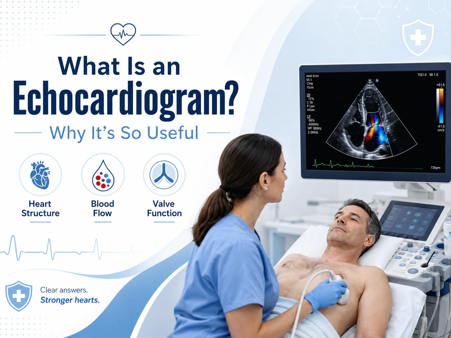
What Is an Echocardiogram and Why Is It So Useful?
An echocardiogram is the medical term for a diagnostic ultrasound procedure performed on the heart. The examination often is referred to as an “echo.” There are more specialized variations of echocardiography, but the standard type is called a transthoracic echocardiogram (TTE). In this test, a sonographer moves a transducer (a device that emits and receives ultrasound waves) across the chest, obtaining images of the heart from different angles. The resulting pictures can help a doctor identify or evaluate cardiac abnormalities.
For a more in-depth discussion of the technology, please see the Wikipedia article on echocardiography. Briefly, this procedure can make use of a related technique called Doppler imaging, which allows visualization of blood flow. Doppler technology can help a physician estimate pressures in various parts of the cardiovascular system and evaluate the function of the heart valves.
A primary value of an echocardiogram lies in its ability to provide anatomical and physiological information about the heart. By reviewing these images, a physician can obtain valuable information about the size and shape of the heart, abnormal movement of the heart muscles, malfunction of the heart valves and other relevant findings. In many cases, an echocardiogram can complement a physical examination, providing insight that might not be apparent from the history or basic tests such as an ECG. A family physician can help determine whether the test is appropriate, and a family physician in Etobicoke can review your blood pressure, cholesterol, diabetes status and other risk factors to help estimate your cardiovascular risk. If the test is indicated, you may receive timely care, as a Markland Wood Medical Clinic representative explains on its website.
What Can Echocardiography Help Doctors Diagnose?
An echocardiogram most commonly is used to evaluate the heart’s pumping function. The heart’s left ventricle is responsible for circulating oxygenated blood to the rest of the body, and an assessment of its function can help identify the presence and severity of heart failure, a condition in which the heart is unable to meet the body’s circulatory demands. Echocardiography often is used when a patient is suspected to have shortness of breath due to fluid overload or diminished pump function.
Heart valve problems also can be diagnosed or evaluated with an echocardiogram. The heart contains four valves: the aortic valve, pulmonary valve, mitral valve and tricuspid valve. These valves control the direction of blood flow through the heart, and echocardiography can help identify improper function, such as narrowing of the valve (stenosis) or backflow of blood (regurgitation). An abnormal heart murmur also can be evaluated with this test, which can help a physician determine whether the finding is benign or suspicious for valve dysfunction.
Echocardiography can help diagnose or assess the extent of other cardiac conditions as well, including dilation of the heart chambers, abnormal movement of the heart muscle, congenital heart defects, fluid around the heart or arrhythmias. In some situations, an echocardiogram can play a role in evaluating patients with irregular heartbeats (arrhythmias). The American Heart Association notes that this procedure can be useful in certain arrhythmia-related scenarios because some arrhythmias are associated with structural heart disease.
It is important to remember that standard echocardiography is not able to directly depict blockages within the coronary arteries. Therefore, a physician who is concerned about restricted blood flow to the heart may recommend alternative tests, such as a stress echocardiogram or other procedures to evaluate the coronary arteries.
Why Faster and On-Site Cardiac Testing Can Matter
A physician’s diagnostic process often involves a series of steps: A patient presents with symptoms, the doctor conducts an evaluation and may order tests to obtain additional information. If an echocardiogram is performed at a distant facility, additional steps are needed to schedule the test, transport the patient if necessary and review the results. These measures can add days or even weeks to the diagnostic process. However, on-site testing typically involves fewer logistical challenges, helping to minimize delays.
The value of faster cardiac testing extends beyond convenience. If an echocardiogram is indicated, a physician can obtain results sooner, potentially leading to earlier diagnosis and intervention. For example, consider a scenario in which a patient presents with unexplained shortness of breath and abnormal findings on a physical exam. If an echocardiogram is performed, it may demonstrate abnormal heart function or valve insufficiency. This information can enable the referring physician to make decisions about cardiology consultation, additional tests, treatment adjustments or other follow-up.
If a patient develops new symptoms, an initial assessment by a physician is needed to determine the cause and the most appropriate type of investigation. In certain situations, a walk-in clinic can help patients receive preliminary care and determine whether outpatient cardiac testing, hospitalization or another type of intervention is needed. For example, a [walk-in clinic in Etobicoke](https://marklandwood.ca/walk-in.html) can assess patients with new palpitations, dizziness or other worrisome symptoms and discuss the option of an echocardiogram or other tests to assess heart function, if appropriate.
Markland Wood Medical Clinic states on its website that it offers non-invasive echocardiography procedures that are interpreted by specialists as part of its cardiac testing program. The clinic also reports that patients seeking on-site echocardiography for assessment of heart function, murmurs, chest pain or other conditions can expect to receive expeditious access to cardiac diagnostic testing.
Echocardiogram vs ECG vs Holter Monitor
When Should a Physician Consider an Echocardiogram?
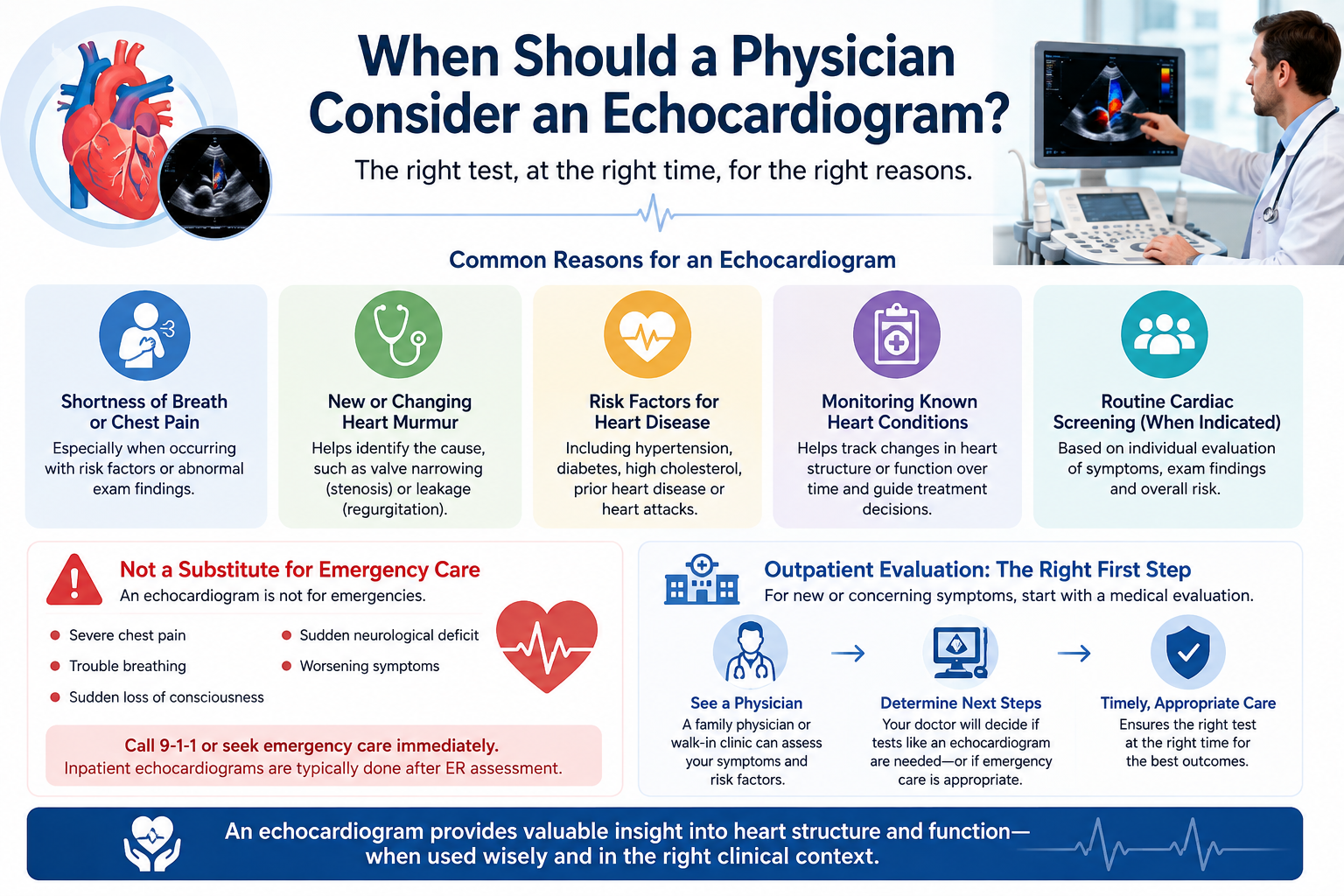
A family physician in Etobicoke can help patients with chronic risk factors receive appropriate cardiac screening. For example, a doctor who sees a patient every year can review recent lab results and blood pressure findings and discuss echocardiography if indicated. By following this approach, the physician can determine whether the test might identify new or evolving abnormalities rather than simply detecting “typical” age-related changes.
Knowing When Outpatient Testing Is Not Enough
Conclusion: Faster Testing Can Create a More Efficient Diagnostic Pathway
Echocardiography is a powerful tool that helps doctors obtain valuable information about the heart. This procedure can help determine whether a patient’s symptoms are related to cardiac dysfunction and provide additional information about the mechanism and severity of disease. When appropriate, on-site echocardiography can help patients avoid some of the delays and hassles associated with diagnostic testing in a distant facility.